
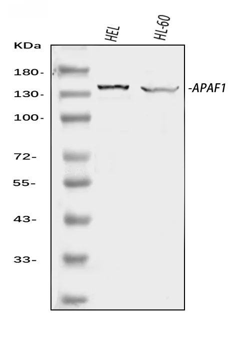
Anti-APAF1 Antibody Picoband®
Kwaliteit
ISO Gecertificeerd
Levering
24-48 uur
Technische Specificaties
Apoptotic peptidase activating factor 1, also known as APAF1, is a protein which in humans is encoded by the APAF1 gene. This gene is mapped to chromosome 12q23. It encodes a cytoplasmic protein that initiates apoptosis. And it is an essential downstream effector of p53-mediated apoptosis. This protein contains several copies of the WD40 repeat domain, a caspase recruitment domain (CARD), and an ATPase domain (NB-ARC) . In the presence of cytochrome c and dATP, APAF1 assembles into an oligomeric apoptosome, which is responsible for activation of procaspase-9 and maintenance of the enzymatic activity of processed caspase-9. Furthermore, APAF1 is inactivated in metastatic melanomas, leading to defects in the execution of apoptotic cell death. Additionally, APAF1 has been shown to interact with NLRP1, Caspase-9, APIP, BCL2-like 1 and HSPA4.
Boster Bio Anti-APAF1 Antibody Picoband® catalog # A00889-2. Tested in ELISA, Flow Cytometry, WB applications. This antibody reacts with Human. The brand Picoband indicates this is a premium antibody that guarantees superior quality, high affinity, and strong signals with minimal background in Western blot applications. Only our best-performing antibodies are designated as Picoband, ensuring unmatched performance.
Zinc finger protein Gfi-1; Growth factor independent protein 1; Zinc finger protein 163; GFI1; ZNF163
APAF1
317
O14727
• Rabbit
Human
No cross-reactivity with other proteins.
E.coli-derived human APAF1 recombinant protein (Position: M1-E1248) .
• Polyclonal
Isoform 1 is expressed in pancreas. Isoform 2 and isoform 3 is expressed in liver.
WB, Flow Cytometry, ELISA
Cancer Susceptibility, Epigenetics and Nuclear Signaling, Proto-Oncogenes, Transcription
Immunogen affinity purified.
Adding 0.2 ml of distilled water will yield a concentration of 500 μg/ml.
Lyophilized
Adding 0.2 ml of distilled water will yield a concentration of 500 μg/ml.
Transcription repressor essential for hematopoiesis. Functions in a cell-context and development-specific manner. Binds to 5'-TAAATCAC[AT]GCA-3' in the promoter region of a large number of genes. Component of several complexes, including the EHMT2-GFI1-HDAC1, AJUBA-GFI1-HDAC1 and RCOR-GFI-KDM1A-HDAC complexes, that suppress, via histone deacetylase (HDAC) recruitment, a number of genes implicated in multilineage blood cell development. Regulates neutrophil differentiation, promotes proliferation of lymphoid cells, and is required for granulocyte development. Mediates, together with U2AF1L4, the alternative splicing of CD45 and controls T-cell receptor signaling. Regulates the endotoxin-mediated Toll-like receptor (TLR) inflammatory response by antagonizing RELA. Cooperates with CBFA2T2 to regulate ITGB1-dependent neurite growth. Controls cell-cycle progression by repressing CDKNIA/p21 transcription in response to TGFB1 via recruitment of GFI1 by ZBTB17 to the CDKNIA/p21 and CDKNIB promoters. Required for the maintenance of inner ear hair cells.
1. Kim, H.; Jung, Y. K.; Kwon, Y. K.; Park, S. H. : Assignment of apoptotic protease activating factor-1 gene (APAF1) to human chromosome band 12q23 by fluorescence in situ hybridization. Cytogenet. Cell Genet. 87: 252-253, 1999. 2. Robles, A. I.; Bemmels, N. A.; Foraker, A. B.; Harris, C. C. : APAF-1 is a transcriptional target of p53 in DNA damage-induced apoptosis. Cancer Res. 61: 6660-6664, 2001. 3. Bao, Q.; Lu, W.; Rabinowitz, J. D.; Shi, Y. : Calcium blocks formation of apoptosome by preventing nucleotide exchange in Apaf-1. Molec. Cell 25: 181-192, 2007. 4. Soengas, M. S.; Capodieci, P.; Polsky, D.; Mora, J.; Esteller, M.; Opitz-Araya, X.; McCombie, R.; Herman, J. G.; Gerald, W. L.; Lazebnik, Y. A.; Cordon-Cardo, C.; Lowe, S. W. : Inactivation of the apoptosis effector Apaf-1 in malignant melanoma. Nature 409: 207-211, 2001. 5. Chu, Z L; Pio F, Xie Z, Welsh K, Krajewska M, Krajewski S, Godzik A, Reed J C (Mar. 2001) . A novel enhancer of the Apaf1 apoptosome involved in cytochrome c-dependent caspase activation and apoptosis. J. Biol. Chem. (United States) 276 (12) : 9239-45.
At -20°C for one year from date of receipt. After reconstitution, at 4°C for one month. It can also be aliquotted and stored frozen at -20°C for six months. Avoid repeated freezing and thawing.
https://www.bosterbio.com/msds?sku=A00889-2
135 kDa
6
Growth factor independent 1 transcriptional repressor
Nucleus.
Rabbit IgG
Each vial contains 4 mg Trehalose, 0.9 mg NaCl, 0.2 mg Na2HPO4.
Beschrijving
Boster Bio Anti-APAF1 Antibody Picoband® catalog # A00889-2. Tested in ELISA, Flow Cytometry, WB applications. This antibody reacts with Human. The brand Pi
Specificaties
Recent bekeken producten

Recombinant TAFV GP1 Protein, C-Fc
EVV03609
100 μg
Gabbr1 Mouse Monoclonal Antibody [Clone ID: S93A-49]
TA326530
100 µg
BSA-Myc/DDK Control Protein
AR100025
50 µL
NH-6
ABC-TC0826
1 Vial
Anti-TAT (Thrombin-antithrombin) Antibody
ABS 014-22-1
1000 µL
Anti-APOE2 mouse monoclonal antibody
TD-AB0132
100 µg
CD35 (CR1) mouse monoclonal antibody, clone J3D3, Azide Free
BM2364
0.2 mg
Anti-KIR2DL1 (Rabbit, Monoclonal, Rabbit mAb)
A115392
100 µL
Recent gezochte producten
Recombinant TAFV GP1 Protein, C-Fc
EVV03609
100 μg
Fluorescein Conjugated Affinity Purified Anti-MOUSE IgG + IgM (H&L) (GOAT) Minimum Cross Reactivity to Bovine, Horse and Human S
GWB-5CB439
1 mg
Horse Polyclonal Horse anti-Goat IgG ImmPRESS(TM) Secondary Antibody [HRP Polymer]
MP-7405-NB-50mL
50 mL
Horse Polyclonal Horse anti-Rabbit IgG ImmPRESS(TM) Secondary Antibody [HRP Polymer]
MP-7401-NB-15mL
15 mL
Horse Polyclonal Horse anti-Mouse IgG ImmPRESS(TM) Secondary Antibody [AP Polymer]
MP-5402-NB-50mL
50 mL
Horse Polyclonal Horse anti-Goat IgG ImmPRESS(TM) Secondary Antibody [HRP Polymer]
MP-7405-NB-15mL
15 mL
Horse Polyclonal Horse anti-Mouse ImmPRESS(TM) VR Secondary Antibody [HRP Polymer]
MP-6402-NB
15 mL
Horse Polyclonal Horse anti-Mouse IgG ImmPRESS(TM) Secondary Antibody [AP Polymer]
MP-5402-NB-15mL
15 mL