
Anti-FAK PTK2 Konijn Monoklonale Antilichaam
Kwaliteit
ISO Gecertificeerd
Levering
24-48 uur
Technische Specificaties
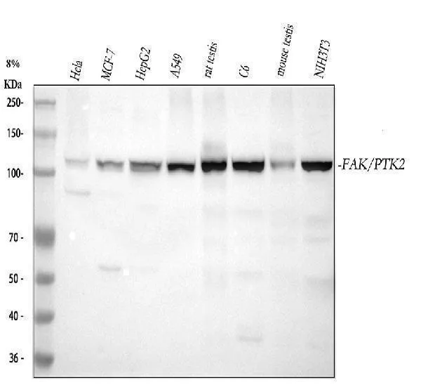
Boster Bio Anti-FAK PTK2 Rabbit Monoclonal Antibody catalog # M00151. Tested in WB, IHC, ICC/IF, IP, Flow Cytometry applications. This antibody reacts with Human, Mouse, Rat.
Focal adhesion kinase 1; FADK 1;2.7.10.2; Focal adhesion kinase-related nonkinase; FRNK; Protein phosphatase 1 regulatory subunit 71; PPP1R71; Protein-tyrosine kinase 2; p125FAK; pp125FAK; PTK2; FAK, FAK1
PTK2
5747
Q05397
• Rabbit
Human, Mouse, Rat
A synthesized peptide derived from human FAK
• Monoclonal
Clone: DDD-16
Detected in B and T-lymphocytes. Isoform 1 and isoform 6 are detected in lung fibroblasts (at protein level) . Ubiquitous. .
WB, IHC, ICC, IF, IP, Flow Cytometry
Angiogenesis, Cancer, Cardiovascular, Cytoskeleton/ECM, Extracellular Matrix, Integrins, Protein Phosphorylation, Signal Transduction, Structures, Tyrosine Kinases
Affinity-chromatography
0.5mg/ml
Liquid
Non-receptor protein-tyrosine kinase that plays an essential role in regulating cell migration, adhesion, spreading, reorganization of the actin cytoskeleton, formation and disassembly of focal adhesions and cell protrusions, cell cycle progression, cell proliferation and apoptosis. Required for early embryonic development and placenta development. Required for embryonic angiogenesis, normal cardiomyocyte migration and proliferation, and normal heart development. Regulates axon growth and neuronal cell migration, axon branching and synapse formation; required for normal development of the nervous system. Plays a role in osteogenesis and differentiation of osteoblasts. Functions in integrin signal transduction, but also in signaling downstream of numerous growth factor receptors, G-protein coupled receptors (GPCR), EPHA2, netrin receptors and LDL receptors. Forms multisubunit signaling complexes with SRC and SRC family members upon activation; this leads to the phosphorylation of additional tyrosine residues, creating binding sites for scaffold proteins, effectors and substrates. Regulates numerous signaling pathways. Promotes activation of phosphatidylinositol 3-kinase and the AKT1 signaling cascade. Promotes activation of MAPK1/ERK2, MAPK3/ERK1 and the MAP kinase signaling cascade. Promotes localized and transient activation of guanine nucleotide exchange factors (GEFs) and GTPase-activating proteins (GAPs), and thereby modulates the activity of Rho family GTPases. Signaling via CAS family members mediates activation of RAC1. Recruits the ubiquitin ligase MDM2 to P53/TP53 in the nucleus, and thereby regulates P53/TP53 activity, P53/TP53 ubiquitination and proteasomal degradation. Phosphorylates SRC; this increases SRC kinase activity. Phosphorylates ACTN1, ARHGEF7, GRB7, RET and WASL. Promotes phosphorylation of PXN and STAT1; most likely PXN and STAT1 are phosphorylated by a SRC family kinase that is recruited to autophosphorylated PTK2/FAK1, rather than by PTK2/FAK1 itself. Promotes phosphorylation of BCAR1; GIT2 and SHC1; this requires both SRC and PTK2/FAK1. Promotes phosphorylation of BMX and PIK3R1. Isoform 6 (FRNK) does not contain a kinase domain and inhibits PTK2/FAK1 phosphorylation and signaling. Its enhanced expression can attenuate the nuclear accumulation of LPXN and limit its ability to enhance serum response factor (SRF) -dependent gene transcription. .
Store at -20°C for one year. For short term storage and frequent use, store at 4°C for up to one month. Avoid repeated freeze-thaw cycles.
https://www.bosterbio.com/msds?sku=M00151
119233 MW
119 kDa
Focal adhesion kinase 1
Cell junction, focal adhesion. Cell membrane; Peripheral membrane protein; Cytoplasmic side. Cytoplasm, cell cortex. Cytoplasm, cytoskeleton. Cytoplasm, cytoskeleton, microtubule organizing center, centrosome . Nucleus. Constituent of focal adhesions. Detected at microtubules.
P2X purinoceptor 1
Rabbit IgG
Rabbit IgG in phosphate buffered saline, pH 7.4, 150mM NaCl, 0.02% sodium azide and 50% glycerol, 0.4-0.5mg/ml BSA.
Beschrijving
Boster Bio Anti-FAK PTK2 Rabbit Monoclonal Antibody catalog # M00151. Tested in WB, IHC, ICC/IF, IP, Flow Cytometry applications. This antibody reacts with Huma
Specificaties
Recent bekeken producten

Recombinant TAFV GP1 Protein, C-Fc
EVV03609
100 μg
Gabbr1 Mouse Monoclonal Antibody [Clone ID: S93A-49]
TA326530
100 µg
BSA-Myc/DDK Control Protein
AR100025
50 µL
NH-6
ABC-TC0826
1 Vial
Anti-TAT (Thrombin-antithrombin) Antibody
ABS 014-22-1
1000 µL
Anti-APOE2 mouse monoclonal antibody
TD-AB0132
100 µg
CD35 (CR1) mouse monoclonal antibody, clone J3D3, Azide Free
BM2364
0.2 mg
Anti-KIR2DL1 (Rabbit, Monoclonal, Rabbit mAb)
A115392
100 µL
Recent gezochte producten
Standard UV Quartz Cell With Lid, 45 X 12.5 X 7.5 mm, Inside Width: 10mm, 1.7ml
SQ003
1 UNIT
Semi-Micro UV Quartz Cell With Frosted Walls and with Teflon Stopper
SQ294
1 UNIT
LoxP Reporter Cell Lines: LoxP-RFP Human Fibroblast (1 x 10⁶ cells, Culture)
ABP-RP-CRFPLOXC
1 flask
Neural precursor cell expressed developmentally down-regulated 8 (NEDD8) [C-terminus] polyclonal antibody
ABP-PAB-10327
100 ug
Neural precursor cell expressed developmentally down-regulated 8 (NEDD8) [N-terminus] polyclonal antibody
ABP-PAB-10326
100 ug
B-cell CLL/lymphoma 10 (BCL10) polyclonal antibody
ABP-PAB-10129
100 ug
LoxP Reporter Cell Lines: loxP-lacZ Cell Line
ABP-RP-CLACLOXE
1 vial
Glial cell derived neurotrophic factor (GDNF) polyclonal antibody
ABP-PAB-10125
100 ug