
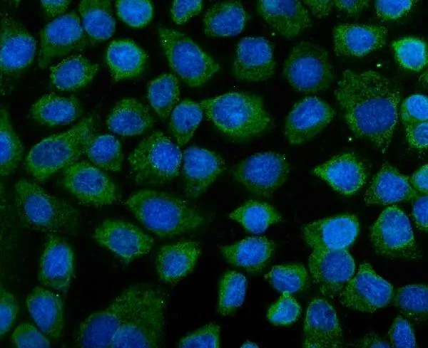
Anti-KEAP1 Antibody Picoband®
Kwaliteit
ISO Gecertificeerd
Levering
24-48 uur
Technische Specificaties
KEAP1 (KELCH-LIKE ECH-ASSOCIATED PROTEIN 1), is a protein that in humans is encoded by the Keap1 gene. The KIAA0132 gene is mapped on 19p13.2. Keap1 contains a central BTB/POZ domain and a C-terminal double glycine repeat (DGR), or Kelch, module. Keap1 has been shown to interact with Nrf2, a master regulator of the antioxidant response, which is important for the amelioration of oxidative stress. In the presence of the electrophilic agent diethylmalate, Nrf2 activity is released from Keap1 and Nrf2 translocate to the nucleus. Under quiescent conditions, Nrf2 is anchored in the cytoplasm through binding to Keap1, which, in turn, facilitates the ubiquitination and subsequent proteolysis of Nrf2. Because Nrf2 activation leads to a coordinated antioxidant and anti-inflammatory response, and Keap1 represses Nrf2 activation, Keap1 has become a very attractive drug target.
Boster Bio Anti-KEAP1 Antibody Picoband® catalog # A00514-3. Tested in ELISA, Flow Cytometry, IF, ICC, WB applications. This antibody reacts with Human, Mouse, Rat. The brand Picoband indicates this is a premium antibody that guarantees superior quality, high affinity, and strong signals with minimal background in Western blot applications. Only our best-performing antibodies are designated as Picoband, ensuring unmatched performance.
Cyclin-dependent-like kinase 5; Cell division protein kinase 5; Serine/threonine-protein kinase PSSALRE; Tau protein kinase II catalytic subunit; TPKII catalytic subunit; CDK5; CDKN5
KEAP1
9817
Q14145
• Rabbit
Human, Mouse, Rat
No cross-reactivity with other proteins.
E.coli-derived human KEAP1 recombinant protein (Position: K84-K312) .
• Polyclonal
Isoform 1 is ubiquitously expressed. Accumulates in cortical neurons (at protein level) . Isoform 2 has only been detected in testis, skeletal muscle, colon, bone marrow and ovary.
WB, ICC, IF, Flow Cytometry, ELISA
Cofactors, Vitamins/Minerals, Hematopoietic Progenitors, Metabolism, Myeloid, Pathways and Processes, Signal Transduction, Stem Cells, Vitamins/Minerals
Immunogen affinity purified.
Adding 0.2 ml of distilled water will yield a concentration of 500 μg/ml.
Lyophilized
Adding 0.2 ml of distilled water will yield a concentration of 500 μg/ml.
Proline-ed serine/threonine-protein kinase essential for neuronal cell cycle arrest and differentiation and may be involved in apoptotic cell death in neuronal diseases by triggering abortive cell cycle re-entry. Interacts with D1 and D3-type G1 cyclins. Phosphorylates SRC, NOS3, VIM/vimentin, p35/CDK5R1, MEF2A, SIPA1L1, SH3GLB1, PXN, PAK1, MCAM/MUC18, SEPT5, SYN1, DNM1, AMPH, SYNJ1, CDK16, RAC1, RHOA, CDC42, TONEBP/NFAT5, MAPT/TAU, MAP1B, histone H1, p53/TP53, HDAC1, APEX1, PTK2/FAK1, huntingtin/HTT, ATM, MAP2, NEFH and NEFM. Regulates several neuronal development and physiological processes including neuronal survival, migration and differentiation, axonal and neurite growth, synaptogenesis, oligodendrocyte differentiation, synaptic plasticity and neurotransmission, by phosphorylating key proteins. Activated by interaction with CDK5R1 (p35) and CDK5R2 (p39), especially in post-mitotic neurons, and promotes CDK5R1 (p35) expression in an autostimulation loop. Phosphorylates many downstream substrates such as Rho and Ras family small GTPases (e.g. PAK1, RAC1, RHOA, CDC42) or microtubule-binding proteins (e.g. MAPT/TAU, MAP2, MAP1B), and modulates actin dynamics to regulate neurite growth and/or spine morphogenesis. Phosphorylates also exocytosis associated proteins such as MCAM/MUC18, SEPT5, SYN1, and CDK16/PCTAIRE1 as well as endocytosis associated proteins such as DNM1, AMPH and SYNJ1 at synaptic terminals. In the mature central nervous system (CNS), regulates neurotransmitter movements by phosphorylating substrates associated with neurotransmitter release and synapse plasticity; synaptic vesicle exocytosis, vesicles fusion with the presynaptic membrane, and endocytosis. Promotes cell survival by activating anti-apoptotic proteins BCL2 and STAT3, and negatively regulating of JNK3/MAPK10 activity. Phosphorylation of p53/TP53 in response to genotoxic and oxidative stresses enhances its stabilization by preventing ubiquitin ligase-mediated proteasomal degradation, and induces transactivation of p53/TP53 target genes, thus regulating apoptosis. Phosphorylation of p35/CDK5R1 enhances its stabilization by preventing calpain-mediated proteolysis producing p25/CDK5R1 and avoiding ubiquitin ligase-mediated proteasomal degradation. During aberrant cell-cycle activity and DNA damage, p25/CDK5 activity elicits cell-cycle activity and double-strand DNA breaks that precedes neuronal death by deregulating HDAC1. DNA damage triggered phosphorylation of huntingtin/HTT in nuclei of neurons protects neurons against polyglutamine expansion as well as DNA damage mediated toxicity. Phosphorylation of PXN reduces its interaction with PTK2/FAK1 in matrix-cell focal adhesions (MCFA) during oligodendrocytes (OLs) differentiation. Negative regulator of Wnt/beta-catenin signaling pathway. Activator of the GAIT (IFN-gamma-activated inhibitor of translation) pathway, which suppresses expression of a post-transcriptional regulon of proinflammatory genes in myeloid cells; phosphorylates the linker domain of glutamyl-prolyl tRNA synthetase (EPRS) in a IFN-gamma-dependent manner, the initial event in assembly of the GAIT complex. Phosphorylation of SH3GLB1 is required for autophagy induction in starved neurons. Phosphorylation of TONEBP/NFAT5 in response to osmotic stress mediates its rapid nuclear localization. MEF2 is inactivated by phosphorylation in nucleus in response to neurotoxin, thus leading to neuronal apoptosis. APEX1 AP-endodeoxyribonuclease is repressed by phosphorylation, resulting in accumulation of DNA damage and contributing to neuronal death. NOS3 phosphorylation down regulates NOS3-derived nitrite (NO) levels. SRC phosphorylation mediates its ubiquitin-dependent degradation and thus leads to cytoskeletal reorganization. May regulate endothelial cell migration and angiogenesis via the modulation of lamellipodia formation. Involved in dendritic spine morphogenesis by mediating the EFNA1-EPHA4 signaling. The complex p35/CDK5 participates in the regulation of the circadian clock by modulating the function of CLOCK protein: phosphorylates CLOCK at 'Thr-451' and 'Thr-461' and regulates the transcriptional activity of the CLOCK-ARNTL/BMAL1 heterodimer in association with altered stability and subcellular distribution.
1. Cullinan, Sara B; Zhang Donna, Hannink Mark, Arvisais Edward, Kaufman Randal J, Diehl J Alan (Oct. 2003) . Nrf2 is a PERK substrate and effector of PERK-dependent cell survival. 2. Itoh, K., Wakabayashi, N., Katoh, Y., Ishii, T., Igarashi, K., Engel, J. D., Yamamoto, M. Keap1 represses nuclear activation of antioxidant responsive elements by Nrf2 through binding to the amino-terminal Neh2 domain. Genes Dev. 13: 76-86, 1999. 3. Nagase, T., Seki, N., Tanaka, A., Ishikawa, K., Nomura, N. Prediction of the coding sequences of unidentified human genes. IV. The coding sequences of 40 new genes (KIAA0121-KIAA0160) deduced by analysis of cDNA clones from human cell line KG-1. DNA Res. 2: 167-174, 1995.
At -20°C for one year from date of receipt. After reconstitution, at 4°C for one month. It can also be aliquotted and stored frozen at -20°C for six months. Avoid repeated freezing and thawing.
https://www.bosterbio.com/msds?sku=A00514-3
40108 MW
66-72 kDa
6
Cyclin-dependent kinase 5
Cell membrane. Peripheral membrane protein. Postsynaptic density. Cytoplasm. Perikaryon. Lamellipodium. Growth cone.
Hereditary hemochromatosis protein
Rabbit IgG
Each vial contains 4 mg Trehalose, 0.9 mg NaCl, 0.2 mg Na2HPO4.
Beschrijving
Boster Bio Anti-KEAP1 Antibody Picoband® catalog # A00514-3. Tested in ELISA, Flow Cytometry, IF, ICC, WB applications. This antibody reacts with Human, Mou
Specificaties
Recent bekeken producten

Anti-APOE2 mouse monoclonal antibody
TD-AB0132
100 µg
CD35 (CR1) mouse monoclonal antibody, clone J3D3, Azide Free
BM2364
0.2 mg
Anti-KIR2DL1 (Rabbit, Monoclonal, Rabbit mAb)
A115392
100 µL
Stains-all
03943-5
5 g
Sudan Blue II;Oil Blue 35
TRC-S688913-500MG
500 mg
Nile Blue A
N8430-01
1 g
Human Apolipoprotein E2 (Apo E2) Antibody
22210-05011
150 µg
MycoBlue Mycoplasma Detector
D101-02
50 Reactions
Recent gezochte producten
Rabbit RBP4 (Retinol Binding Protein 4) ELISA Kit
ERB0177
96 Tests
Rabbit RBP ELISA Kit
ERTR0013
96 Tests
Rabbit RBP4 ELISA Kit
ERTR0008
96 Tests
Rabbit RBP4 (Retinol Binding Protein 4, Plasma) ELISA Kit
EK251411
96 Well
GENLISA™ Rabbit Retinol Binding Protein 4 Kit (Rabbit RBP4) ELISA
KLX0480
1x 96 Well
Rabbit RBP4 (Retinol Binding Protein 4) ELISA Kit
MBS7612483-01
48 Well
Rabbit RBP4 ELISA Kit
E095570
1 Each
Rabbit RBP5 ELISA Kit
E109374
1 Each