
Anti-Niemann Pick C1 Konijn Monoklonale Antilichaam
Kwaliteit
ISO Gecertificeerd
Levering
24-48 uur
Technische Specificaties
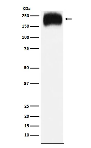
Boster Bio Anti-Niemann Pick C1 Rabbit Monoclonal Antibody catalog # M00428-1. Tested in WB, IHC, ICC/IF, Flow Cytometry applications. This antibody reacts with Human, Mouse, Rat.
Prolyl endopeptidase FAP ;3.4.21.26 ;170 kDa melanoma membrane-bound gelatinase ; Dipeptidyl peptidase FAP ;3.4.14.5 ; Fibroblast activation protein alpha ; FAPalpha ; Gelatine degradation protease FAP ;3.4.21.- ; Integral membrane serine protease ; Post-proline cleaving enzyme ; Serine integral membrane protease ; SIMP ; Surface-expressed protease ; Seprase ; Antiplasmin-cleaving enzyme FAP, soluble form ; APCE ;3.4.14.5 ;3.4.21.- ;3.4.21.26 ; FAP
NPC1
4864
O15118
• Rabbit
Human, Mouse, Rat
A synthesized peptide derived from human Niemann Pick C1
• Monoclonal
Clone: 18N07
Expressed in adipose tissue. Expressed in the dermal fibroblasts in the fetal skin. Expressed in the granulation tissue of healing wounds and on reactive stromal fibroblast in epithelial cancers. Expressed in activated fibroblast-like synoviocytes from inflamed synovial tissues. Expressed in activated hepatic stellate cells (HSC) and myofibroblasts from cirrhotic liver, but not detected in normal liver. Expressed in glioma cells (at protein level) . Expressed in glioblastomas and glioma cells. Isoform 1 and isoform 2 are expressed in melanoma, carcinoma and fibroblast cell lines. .
WB, IHC, ICC, IF, Flow Cytometry
Adaptive Immunity, Immunoglobulins, Immunology, Protein Phosphorylation, Receptors, Secreted Molecules, Signal Transduction, T Cells, Tyrosine Kinases
Affinity-chromatography
0.5mg/ml
Liquid
Cell surface glycoprotein serine protease that participates in extracellular matrix degradation and involved in many cellular processes including tissue remodeling, fibrosis, wound healing, inflammation and tumor growth. Both plasma membrane and soluble forms exhibit post-proline cleaving endopeptidase activity, with a marked preference for Ala/Ser-Gly-Pro-Ser/Asn/Ala consensus sequences, on substrate such as alpha-2-antiplasmin SERPINF2 and SPRY2 (PubMed:14751930, PubMed:16223769, PubMed:16480718, PubMed:16410248, PubMed:17381073, PubMed:18095711, PubMed:21288888, PubMed:24371721) . Degrade also gelatin, heat-denatured type I collagen, but not native collagen type I and IV, vibronectin, tenascin, laminin, fibronectin, fibrin or casein (PubMed:9065413, PubMed:2172980, PubMed:7923219, PubMed:10347120, PubMed:10455171, PubMed:12376466, PubMed:16223769, PubMed:16651416, PubMed:18095711) . Have also dipeptidyl peptidase activity, exhibiting the ability to hydrolyze the prolyl bond two residues from the N-terminus of synthetic dipeptide substrates provided that the penultimate residue is proline, with a preference for Ala-Pro, Ile-Pro, Gly-Pro, Arg-Pro and Pro-Pro (PubMed:10347120, PubMed:10593948, PubMed:16175601, PubMed:16223769, PubMed:16651416, PubMed:16410248, PubMed:17381073, PubMed:21314817, PubMed:24371721, PubMed:24717288) . Natural neuropeptide hormones for dipeptidyl peptidase are the neuropeptide Y (NPY), peptide YY (PYY), substance P (TAC1) and brain natriuretic peptide 32 (NPPB) (PubMed:21314817) . The plasma membrane form, in association with either DPP4, PLAUR or integrins, is involved in the pericellular proteolysis of the extracellular matrix (ECM), and hence promotes cell adhesion, migration and invasion through the ECM. Plays a role in tissue remodeling during development and wound healing. Participates in the cell invasiveness towards the ECM in malignant melanoma cancers. Enhances tumor growth progression by increasing angiogenesis, collagen fiber degradation and apoptosis and by reducing antitumor response of the immune system. Promotes glioma cell invasion through the brain parenchyma by degrading the proteoglycan brevican. Acts as a tumor suppressor in melanocytic cells through regulation of cell proliferation and survival in a serine protease activity-independent manner. .
Store at -20°C for one year. For short term storage and frequent use, store at 4°C for up to one month. Avoid repeated freeze-thaw cycles.
https://www.bosterbio.com/msds?sku=M00428-1
87713 MW
160-180 kDa
Prolyl endopeptidase FAP
Prolyl endopeptidase FAP: Cell surface . Cell membrane ; Single- pass type II membrane protein . Cell projection, lamellipodium membrane ; Single-pass type II membrane protein . Cell projection, invadopodium membrane ; Single-pass type II membrane protein . Cell projection, ruffle membrane ; Single-pass type II membrane protein . Membrane ; Single-pass type II membrane protein . Localized on cell surface with lamellipodia and invadopodia membranes and on shed vesicles. Colocalized with DPP4 at invadopodia and lamellipodia membranes of migratory activated endothelial cells in collagenous matrix. Colocalized with DPP4 on endothelial cells of capillary-like microvessels but not large vessels within invasive breast ductal carcinoma. Anchored and enriched preferentially by integrin alpha-3/beta-1 at invadopodia, plasma membrane protrusions that correspond to sites of cell invasion, in a collagen-dependent manner. Localized at plasma and ruffle membranes in a collagen-independent manner. Colocalized with PLAUR preferentially at the cell surface of invadopodia membranes in a cytoskeleton-, integrin- and vitronectin-dependent manner. Concentrated at invadopodia membranes, specialized protrusions of the ventral plasma membrane in a fibrobectin-dependent manner. Colocalizes with extracellular components (ECM), such as collagen fibers and fibronectin. .
T-cell surface glycoprotein CD4
IgG
Rabbit IgG in phosphate buffered saline, pH 7.4, 150mM NaCl, 0.02% sodium azide and 50% glycerol, 0.4-0.5mg/ml BSA.
Beschrijving
Boster Bio Anti-Niemann Pick C1 Rabbit Monoclonal Antibody catalog # M00428-1. Tested in WB, IHC, ICC/IF, Flow Cytometry applications. This antibody reacts with
Specificaties
Recent bekeken producten

MARV VP40/Marburg VP40 Protein (N-His, Lake Victoria marburgvirus (strain Musoke-80) (MARV) (Marburg virus (strain Kenya/Musoke/1980) ) )
P505862
100 µg
Hettich Centrifuge Rotanta 460R Refrigerated Large Volume
CEN0703
1 Each
X4RF Refrigerated Centrifuge
75009936
1 Each
Drug Stability Chamber
500-DSC102
1 Unit
Modular Incubator Chamber
MIC-101
1 Unit
CYP707A2 Antibody
GTR17947107
2 mg

SsDNA Assay Kit
12645ES60
100 Tests
Eco Alu MTP (HxD) 6x7=42 plates 35mm, 223x630x135mm
TE15336
1 Piece
Recent gezochte producten
MARV VP40/Marburg VP40 Protein (N-His, Lake Victoria marburgvirus (strain Musoke-80) (MARV) (Marburg virus (strain Kenya/Musoke/1980) ) )
P505862
100 µg
X4RF Refrigerated Centrifuge
75009936
1 Each
Low Speed Refrigerated Centrifuge
LX104LSR
1 Unit
Low Speed Refrigerated Centrifuge
LX117LSR
1 Unit
Low Speed Refrigerated Centrifuge
LX124LSR
1 Unit
Blood Bank Refrigerator
LX4000BBR
1 Unit
Blood Bank Refrigerator
LX4002BBR
1 Unit
Blood Bank Refrigerator
LX4003BBR
1 Unit