
Anti-Prolactin Receptor Konijn Monoklonale Antilichaam
Kwaliteit
ISO Gecertificeerd
Levering
24-48 uur
Technische Specificaties
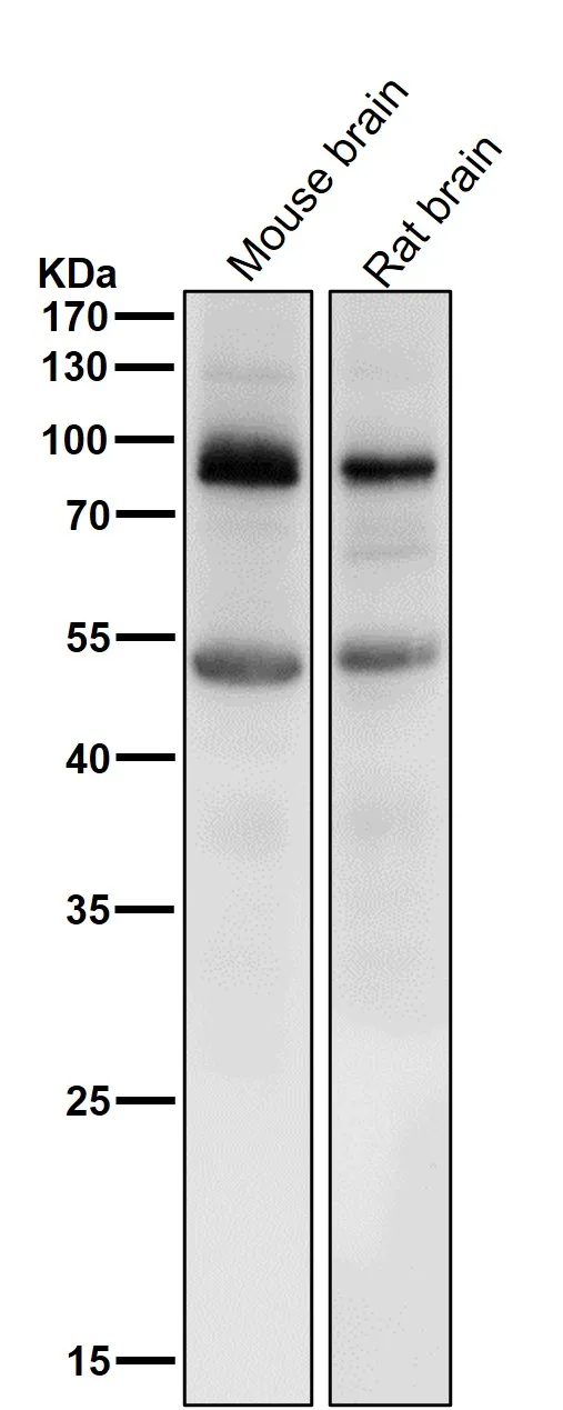
Boster Bio Anti-Prolactin Receptor Rabbit Monoclonal Antibody catalog # M01196-2. Tested in WB, IHC, ICC/IF applications. This antibody reacts with Human, Mouse, Rat.
HLA class II histocompatibility antigen, DR alpha chain; MHC class II antigen DRA; HLA-DRA; HLA-DRA1
PRLR
5618
P16471
• Rabbit
Human, Mouse, Rat
A synthesized peptide derived from human Prolactin Receptor.
• Monoclonal
Clone: ACIF-16
Ubiquitous. Expression is strongest in fetal and adult brain, spinal cord, and cerebellum, followed by thymus, bone marrow, testis, and fetal liver. Expression is intermediate in colon, ovary, placenta, uterus, and trachea, and is readily detected at substantially lower levels in all other tissues examined. Lowest expression is found in adult liver. Present in much greater abundance in cells from patients with acute leukemia of different subtypes than in normal peripheral blood lymphocytes, non-leukemic proliferating lymphoid cells, bone marrow cells, or cells from patients with chronic lymphoid or myeloid leukemia. .
WB, IHC, ICC, IF
Cell Biology, Cell Cycle, Cell Differentiation, Growth Factors/Hormones, Signal Transduction
Affinity-chromatography
0.5mg/ml
Liquid
Binds peptides derived from antigens that access the endocytic route of antigen presenting cells (APC) and presents them on the cell surface for recognition by the CD4 T-cells. The peptide binding cleft accommodates peptides of 10-30 residues. The peptides presented by MHC class II molecules are generated mostly by degradation of proteins that access the endocytic route, where they are processed by lysosomal proteases and other hydrolases. Exogenous antigens that have been endocytosed by the APC are thus readily available for presentation via MHC II molecules, and for this reason this antigen presentation pathway is usually referred to as exogenous. As membrane proteins on their way to degradation in lysosomes as part of their normal turn-over are also contained in the endosomal/lysosomal compartments, exogenous antigens must compete with those derived from endogenous components. Autophagy is also a source of endogenous peptides, autophagosomes constitutively fuse with MHC class II loading compartments. In addition to APCs, other cells of the gastrointestinal tract, such as epithelial cells, express MHC class II molecules and CD74 and act as APCs, which is an unusual trait of the GI tract. To produce a MHC class II molecule that presents an antigen, three MHC class II molecules (heterodimers of an alpha and a beta chain) associate with a CD74 trimer in the ER to form a heterononamer. Soon after the entry of this complex into the endosomal/lysosomal system where antigen processing occurs, CD74 undergoes a sequential degradation by various proteases, including CTSS and CTSL, leaving a small fragment termed CLIP (class-II-associated invariant chain peptide) . The removal of CLIP is facilitated by HLA-DM via binding to the alpha-beta-CLIP complex so that CLIP is released. HLA-DM stabilizes MHC class II molecules until primary high affinity antigenic peptides are bound. The MHC II molecule bound to a peptide is then transported to the cell membrane surface. In B-cells, the interaction between HLA-DM and MHC class II molecules is regulated by HLA-DO. Primary dendritic cells (DCs) also to express HLA-DO. Lysosomal microenvironment has been implicated in the regulation of antigen loading into MHC II molecules, increased acidification produces increased proteolysis and efficient peptide loading.
Store at -20°C for one year. For short term storage and frequent use, store at 4°C for up to one month. Avoid repeated freeze-thaw cycles.
https://www.bosterbio.com/msds?sku=M01196-2
28607 MW
17 kDa
HLA class II histocompatibility antigen, DR alpha chain
Cell membrane; Single-pass type I membrane protein. Endoplasmic reticulum membrane; Single-pass type I membrane protein. Golgi apparatus, trans-Golgi network membrane; Single-pass type I membrane protein. Endosome membrane; Single- pass type I membrane protein. Lysosome membrane; Single-pass type I membrane protein. Late endosome membrane; Single-pass type I membrane protein. The MHC class II complex transits through a number of intracellular compartments in the endocytic pathway until it reaches the cell membrane for antigen presentation.
Glycogen synthase kinase-3 beta (gsk3b)
Rabbit IgG
Rabbit IgG in phosphate buffered saline, pH 7.4, 150mM NaCl, 0.02% sodium azide and 50% glycerol, 0.4-0.5mg/ml BSA.
Beschrijving
Boster Bio Anti-Prolactin Receptor Rabbit Monoclonal Antibody catalog # M01196-2. Tested in WB, IHC, ICC/IF applications. This antibody reacts with Human, Mouse
Specificaties
Recent bekeken producten

MARV VP40/Marburg VP40 Protein (N-His, Lake Victoria marburgvirus (strain Musoke-80) (MARV) (Marburg virus (strain Kenya/Musoke/1980) ) )
P505862
100 µg
Hettich Centrifuge Rotanta 460R Refrigerated Large Volume
CEN0703
1 Each
X4RF Refrigerated Centrifuge
75009936
1 Each
Drug Stability Chamber
500-DSC102
1 Unit
Modular Incubator Chamber
MIC-101
1 Unit
CYP707A2 Antibody
GTR17947107
2 mg

SsDNA Assay Kit
12645ES60
100 Tests
Eco Alu MTP (HxD) 6x7=42 plates 35mm, 223x630x135mm
TE15336
1 Piece
Recent gezochte producten
MARV VP40/Marburg VP40 Protein (N-His, Lake Victoria marburgvirus (strain Musoke-80) (MARV) (Marburg virus (strain Kenya/Musoke/1980) ) )
P505862
100 µg
X4RF Refrigerated Centrifuge
75009936
1 Each
Low Speed Refrigerated Centrifuge
LX104LSR
1 Unit
Low Speed Refrigerated Centrifuge
LX117LSR
1 Unit
Low Speed Refrigerated Centrifuge
LX124LSR
1 Unit
Blood Bank Refrigerator
LX4000BBR
1 Unit
Blood Bank Refrigerator
LX4002BBR
1 Unit
Blood Bank Refrigerator
LX4003BBR
1 Unit