
Anti-Rho A + B + C Konijn Monoklonale Antilichaam
Kwaliteit
ISO Gecertificeerd
Levering
24-48 uur
Technische Specificaties
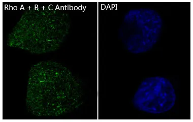
Boster Bio Anti-Rho A + B + C Rabbit Monoclonal Antibody catalog # M00207-1. Tested in WB, IHC, ICC/IF, Flow Cytometry applications. This antibody reacts with Human, Mouse, Rat.
Transforming protein RhoA; Rho cDNA clone 12; h12; RHOA; ARH12, ARHA, RHO12
RHOA
387
P61586/P62745/P08134
• Rabbit
Human, Mouse, Rat
A synthesized peptide derived from human Rho A + B + C
• Monoclonal
Clone: DGF-18
Expressed in brain, kidney and testis. Very low or no expression in spleen, heart, and muscle. In 20- to 22- week old fetuses it is expressed at high level in kidney, lung, small intestine and brain, and to a lower degree in spleen, liver, and muscle. Isoform 2 is detected in epithelial cells. Isoform 1 is not detected in epithelial cells. Isoform 1 and isoform 2 are detected in fibroblastic cells. .
WB, IHC, ICC, IF, Flow Cytometry
Actin Assembly, Actin, etc., Cancer, Cytoskeleton, Cytoskeleton/ECM, G Protein Signaling, Microfilaments, Ras Family, Signal Transduction, Signaling Pathway, Small G Proteins
Affinity-chromatography
0.5mg/ml
Liquid
Regulates a signal transduction pathway linking plasma membrane receptors to the assembly of focal adhesions and actin stress fibers. Involved in a microtubule-dependent signal that is required for the myosin contractile ring formation during cell cycle cytokinesis. Plays an essential role in cleavage furrow formation. Required for the apical junction formation of keratinocyte cell-cell adhesion. Stimulates PKN2 kinase activity. May be an activator of PLCE1. Activated by ARHGEF2, which promotes the exchange of GDP for GTP. Essential for the SPATA13-mediated regulation of cell migration and adhesion assembly and disassembly. The MEMO1-RHOA-DIAPH1 signaling pathway plays an important role in ERBB2-dependent stabilization of microtubules at the cell cortex. It controls the localization of APC and CLASP2 to the cell membrane, via the regulation of GSK3B activity. In turn, membrane-bound APC allows the localization of the MACF1 to the cell membrane, which is required for microtubule capture and stabilization. Regulates a signal transduction pathway linking plasma membrane receptors to the assembly of focal adhesions and actin stress fibers. Involved in a microtubule-dependent signal that is required for the myosin contractile ring formation during cell cycle cytokinesis. Plays an essential role in cleavage furrow formation. Required for the apical junction formation of keratinocyte cell-cell adhesion. May be an activator of PLCE1. Activated by ARHGEF2, which promotes the exchange of GDP for GTP. Essential for the SPATA13-mediated regulation of cell migration and adhesion assembly and disassembly. The MEMO1-RHOA-DIAPH1 signaling pathway plays an important role in ERBB2-dependent stabilization of microtubules at the cell cortex. It controls the localization of APC and CLASP2 to the cell membrane, via the regulation of GSK3B activity. In turn, membrane-bound APC allows the localization of the MACF1 to the cell membrane, which is required for microtubule capture and stabilization (By similarity) . Regulates KCNA2 potassium channel activity by reducing its location at the cell surface in response to CHRM1 activation; promotes KCNA2 endocytosis (PubMed:9635436, PubMed:19403695) .
Store at -20°C for one year. For short term storage and frequent use, store at 4°C for up to one month. Avoid repeated freeze-thaw cycles.
https://www.bosterbio.com/msds?sku=M00207-1
21768 MW
103 kDa
Transforming protein RhoA
Cell membrane; Lipid-anchor; Cytoplasmic side. Cytoplasm, cytoskeleton. Cleavage furrow. Cytoplasm, cell cortex . Midbody. Cell projection, lamellipodium . Localized to cell-cell contacts in calcium-treated keratinocytes (By similarity) . Translocates to the equatorial region before furrow formation in a ECT2-dependent manner. Localizes to the equatorial cell cortex (at the site of the presumptive furrow) in early anaphase in a activated form and in a myosin- and actin-independent manner. .
P2X purinoceptor 1
Rabbit IgG
Rabbit IgG in phosphate buffered saline, pH 7.4, 150mM NaCl, 0.02% sodium azide and 50% glycerol, 0.4-0.5mg/ml BSA.
Beschrijving
Boster Bio Anti-Rho A + B + C Rabbit Monoclonal Antibody catalog # M00207-1. Tested in WB, IHC, ICC/IF, Flow Cytometry applications. This antibody reacts with H
Specificaties
Recent bekeken producten

Recombinant TAFV GP1 Protein, C-Fc
EVV03609
100 μg
Gabbr1 Mouse Monoclonal Antibody [Clone ID: S93A-49]
TA326530
100 µg
BSA-Myc/DDK Control Protein
AR100025
50 µL
NH-6
ABC-TC0826
1 Vial
Anti-TAT (Thrombin-antithrombin) Antibody
ABS 014-22-1
1000 µL
Anti-APOE2 mouse monoclonal antibody
TD-AB0132
100 µg
CD35 (CR1) mouse monoclonal antibody, clone J3D3, Azide Free
BM2364
0.2 mg
Anti-KIR2DL1 (Rabbit, Monoclonal, Rabbit mAb)
A115392
100 µL
Recent gezochte producten
Recombinant TAFV GP1 Protein, C-Fc
EVV03609
100 μg
Fluorescein Conjugated Affinity Purified Anti-MOUSE IgG + IgM (H&L) (GOAT) Minimum Cross Reactivity to Bovine, Horse and Human S
GWB-5CB439
1 mg
Horse Polyclonal Horse anti-Goat IgG ImmPRESS(TM) Secondary Antibody [HRP Polymer]
MP-7405-NB-50mL
50 mL
Horse Polyclonal Horse anti-Rabbit IgG ImmPRESS(TM) Secondary Antibody [HRP Polymer]
MP-7401-NB-15mL
15 mL
Horse Polyclonal Horse anti-Mouse IgG ImmPRESS(TM) Secondary Antibody [AP Polymer]
MP-5402-NB-50mL
50 mL
Horse Polyclonal Horse anti-Goat IgG ImmPRESS(TM) Secondary Antibody [HRP Polymer]
MP-7405-NB-15mL
15 mL
Horse Polyclonal Horse anti-Mouse ImmPRESS(TM) VR Secondary Antibody [HRP Polymer]
MP-6402-NB
15 mL
Horse Polyclonal Horse anti-Mouse IgG ImmPRESS(TM) Secondary Antibody [AP Polymer]
MP-5402-NB-15mL
15 mL