

Anti-zware ketting Myosine/MYH3 Antilichaam Picoband® fluoro550 Vermengd
Kwaliteit
ISO Gecertificeerd
Levering
24-48 uur
Technische Specificaties
Myosin-3 is a protein that in humans is encoded by the MYH3 gene. Myosin is a major contractile protein which converts chemical energy into mechanical energy through the hydrolysis of ATP. Myosin is a hexameric protein composed of a pair of myosin heavy chains (MYH) and two pairs of nonidentical light chains. This gene is a member of the MYH family and encodes a protein with an IQ domain and a myosin head-like domain. Mutations in this gene have been associated with two congenital contracture (arthrogryposis) syndromes, Freeman-Sheldon syndrome and Sheldon-Hall syndrome.
Neuronal PAS domain-containing protein 2; Neuronal PAS2; Basic-helix-loop-helix-PAS protein MOP4; Class E basic helix-loop-helix protein 9; bHLHe9; Member of PAS protein 4; PAS domain-containing protein 4; NPAS2; BHLHE9, MOP4, PASD4
MYH3
4621
P11055
• Rabbit
Human, Mouse, Rat
No cross-reactivity with other proteins.
E.coli-derived human heavy chain Myosin/MYH3 recombinant protein (Position: K44-E1940) .
• Polyclonal
Flow Cytometry
Atherosclerosis, Cardiovascular, Domain Families, Epigenetics and Nuclear Signaling, Hlh/Leucine Zipper, Transcription, Transcription Factors
Immunogen affinity purified.
Liquid
Transcriptional activator which forms a core component of the circadian clock. The circadian clock, an internal time- keeping system, regulates various physiological processes through the generation of approximately 24 hour circadian rhythms in gene expression, which are translated into rhythms in metabolism and behavior. It is derived from the Latin roots 'circa' (about) and 'diem' (day) and acts as an important regulator of a wide array of physiological functions including metabolism, sleep, body temperature, blood pressure, endocrine, immune, cardiovascular, and renal function. Consists of two major components: the central clock, residing in the suprachiasmatic nucleus (SCN) of the brain, and the peripheral clocks that are present in nearly every tissue and organ system. Both the central and peripheral clocks can be reset by environmental cues, also known as Zeitgebers (German for 'timegivers') . The predominant Zeitgeber for the central clock is light, which is sensed by retina and signals ly to the SCN. The central clock entrains the peripheral clocks through neuronal and hormonal signals, body temperature and feeding-related cues, aligning all clocks with the external light/dark cycle. Circadian rhythms allow an organism to achieve temporal homeostasis with its environment at the molecular level by regulating gene expression to create a peak of protein expression once every 24 hours to control when a particular physiological process is most active with respect to the solar day. Transcription and translation of core clock components (CLOCK, NPAS2, ARNTL/BMAL1, ARNTL2/BMAL2, PER1, PER2, PER3, CRY1 and CRY2) plays a critical role in rhythm generation, whereas delays imposed by post-translational modifications (PTMs) are important for determining the period (tau) of the rhythms (tau refers to the period of a rhythm and is the length, in time, of one complete cycle) . A diurnal rhythm is synchronized with the day/night cycle, while the ultradian and infradian rhythms have a period shorter and longer than 24 hours, respectively. Disruptions in the circadian rhythms contribute to the pathology of cardiovascular diseases, cancer, metabolic syndromes and aging. A transcription/translation feedback loop (TTFL) forms the core of the molecular circadian clock mechanism. Transcription factors, CLOCK or NPAS2 and ARNTL/BMAL1 or ARNTL2/BMAL2, form the positive limb of the feedback loop, act in the form of a heterodimer and activate the transcription of core clock genes and clock-controlled genes (involved in key metabolic processes), harboring E-box elements (5'-CACGTG-3') within their promoters. The core clock genes: PER1/2/3 and CRY1/2 which are transcriptional repressors form the negative limb of the feedback loop and interact with the CLOCK|NPAS2-ARNTL/BMAL1|ARNTL2/BMAL2 heterodimer inhibiting its activity and thereby negatively regulating their own expression. This heterodimer also activates nuclear receptors NR1D1/2 and RORA/B/G, which form a second feedback loop and which activate and repress ARNTL/BMAL1 transcription, respectively. The NPAS2-ARNTL/BMAL1 heterodimer positively regulates the expression of MAOA, F7 and LDHA and modulates the circadian rhythm of daytime contrast sensitivity by regulating the rhythmic expression of adenylate cyclase type 1 (ADCY1) in the retina. NPAS2 plays an important role in sleep homeostasis and in maintaining circadian behaviors in normal light/dark and feeding conditions and in the effective synchronization of feeding behavior with scheduled food availability. Regulates the gene transcription of key metabolic pathways in the liver and is involved in DNA damage response by regulating several cell cycle and DNA repair genes. .
1. Cameron-Christie, S. R., Wells, C. F., Simon, M., Wessels, M., Tang, C. Z. N., Wei, W., Takei, R., Aarts-Tesselaar, C., Sandaradura, S., Sillence, D. O., Cordier, M.-P, , Veenstra-Knol, H. E., Cassina, M., Ludkig, K., Trevission, E., Bahlo, M., Markie, D. M., Jenkins, Z. A., Robertson, S. P. Recessive spondylocarpotarsal synostosis syndrome due to compound heterozygosity for variants in MYH3. Am. J. Hum. Genet. 102: 1115-1125, 2018. Note: Erratum: Am. J. Hum. Genet. 105: 669 only, 2019. 2. Carapito, R., Goldenberg, A., Paul, N., Pichot, A., David, A., Hamel, A., Dumant-Forest, C., Leroux, J., Ory, B., Isidor, B., Bahram, S. Protein-altering MYH3 variants are associated with a spectrum of phenotypes extending to spondylocarpotarsal synostosis syndrome. Europ. J. Hum. Genet. 24: 1746-1751, 2016. 3. Chong, J. X., Burrage, L. C., Beck, A. E., Marvin, C. T., McMillin, M. J., Shively, K. M., Harrell, T. M., Buckingham, K. J., Bacino, C. A., Jain, M., Alanay, Y., Berry, S. A., Carey, J. C., Gibbs, R. A., Lee, B. H., Krakow, D., Shendure, J., Nickerson, D. A., University of Washington Center for Mendelian Genomics, Bamshad, M. J. Autosomal-dominant multiple pterygium syndrome is caused by mutations in MYH3. Am. J. Hum. Genet. 96: 841-849, 2015.
At -20 ̊C for one year from date of receipt. Avoid repeated freezing and thawing. Protect from light.
91791 MW
6
Neuronal PAS domain-containing protein 2
Nucleus .
Rabbit IgG
Each vial contains 50% glycerol, 0.9% NaCl, 0.2% Na2HPO4, 0.02% NaN3.
Beschrijving
Anti-zware ketting Myosine/MYH3 Antilichaam Picoband® fluoro550 Vermengd Beschikbaar in 100 µg/Vial. Bestel eenvoudig online met snelle levering.
Specificaties
Recent bekeken producten
Cellubrevin Antibody
MBS9436639-01
0.05 mL
Oxidative Stress Combo Assay Tissue Kit
EA-7008
1 Each
Arachis hypogaea Lectin (PNA) - Biotinylated
21510020-1
5mg
1,4- Dioxane Pure
1041330 500
500 mL
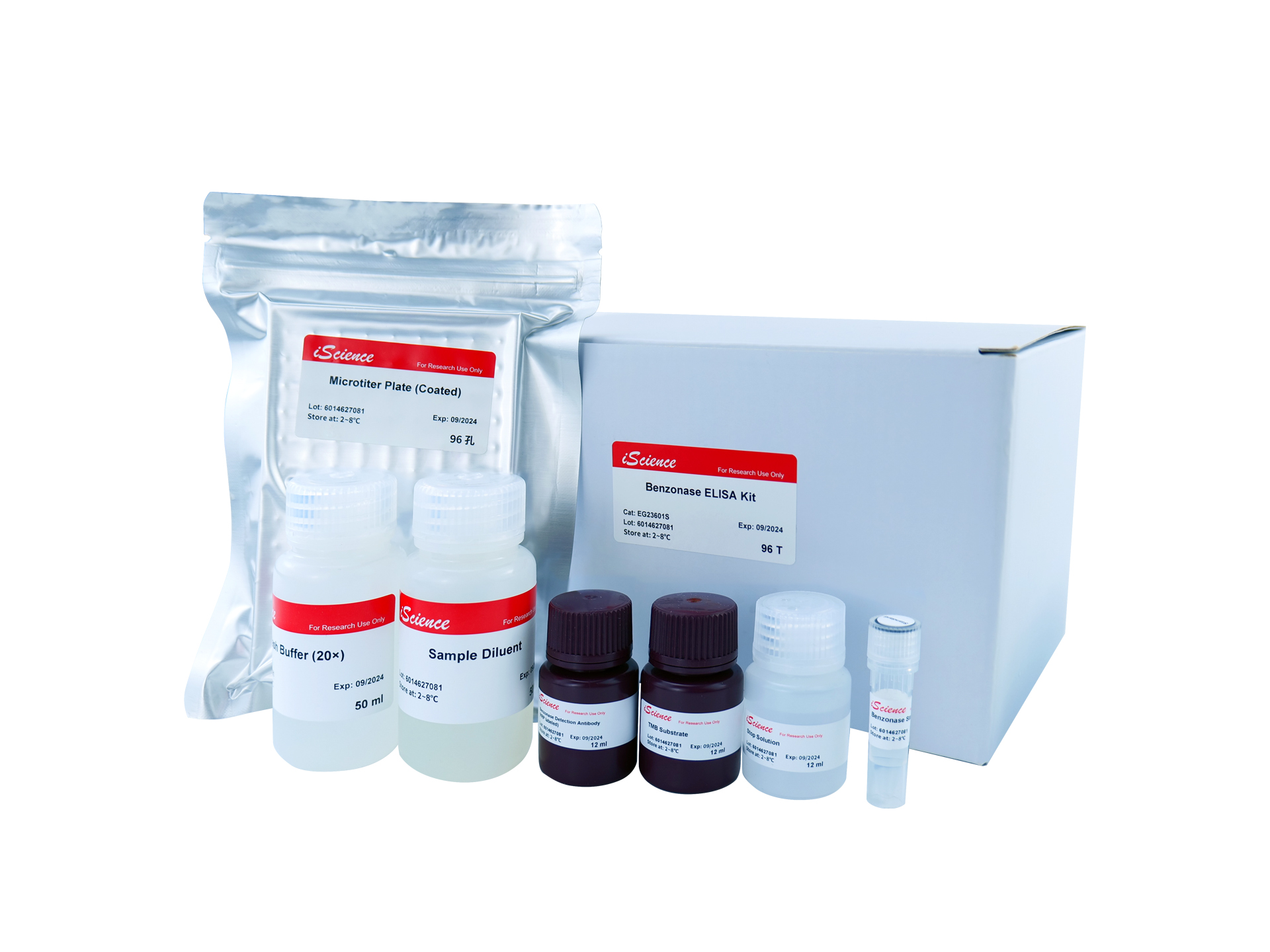
Benzonase ELISA Kit
EG23601S
96 Tests

Not I Restriction Enzyme
B2016623
500 Units
K562-HLA-G0101 Overexpressing Cell Line
RG-1214
5x 10^6
Yeast Poly (A) Polymerase
RNT-006-S
30000 Units
Recent gezochte producten
Cellubrevin Antibody
MBS9436639-01
0.05 mL
Rat Oxidative Stress Primer Library
ROSL-I
1 set
DTNB (Ellman’s) Thiol Oxidative Stress Assay
1021
1 Kit
OSR1 (Oxidative Stress Responsive 1)
MBS616581-01
0.1 mL
Oxidative Stress Combo Assay Cell Kit
EA-7006
1 Each
Oxidative Stress Combo Assay Blood Kit
EA-7007
1 Each
Oxidative Stress Combo Assay Tissue Kit
EA-7008
1 Each
OSR1 (Oxidative Stress Responsive 1)
O8054
100 µL