

Humaan alfa-fetoproteiFP antilichaam
Kwaliteit
ISO Gecertificeerd
Levering
24-48 uur
Technische Specificaties
AFP, also known as alpha-fetoprotein, is a major plasma protein produced by the yolk sac and the liver during fetal development. It is thought to be the fetal form of serum albumin. This protein binds to copper, nickel, fatty acids and bilirubin. The main function of this is to prevent the virilization of female fetuses. As it does not bind estrogen, its function in humans is less clear.
Alpha-fetoprotein, AFPD, FETA, HPAFP
• Mouse
Human
Human
Recombinant human AFP (19-609aa) purified from insect cell.
• Monoclonal
• IgG1 κ
AT73E10
Unconjugated
ELISA, WB, ICC/IF, FACS
By protein-A affinity chromatography
1 mg/mL (determined by BCA assay)
A-Fetoprotein, AFP, ATGA0488-10 µg, ATGA0488-20 µg, ATGA0488-50 µg, ATGA0488-100 µg, ATGA0488-250 µg, ATGA0488-500 µg, ATGA0488-1 mg, ATGA0488-10, ATGA0488-20, ATGA0488-50, ATGA0488-100, ATGA0488-250, ATGA0488-500, ATGA0488-1
Tomasi TB Jr., (1997) Annu Rev Med. 28: 453-465. ; ; Elbrecht A, et al. (1992) Science. 255: 467-470.
Can be stored at 2°C to 8°C for 1 week. For long term storage, aliquot and store at -20C to -80C. Avoid repeated freezing and thawing cycles.
Liquid in. Phosphate-Buffered Saline (pH 7.4) with 0.02% Sodium Azide, 10% glycerol
The antibody has been tested by ELISA, Western blot analysis, Flow cytometry and ICC/IF to assure specificity and reactivity. Since application varies, however, each investigation should be titrated by the reagent to obtain optimal results.
Cardiovascular
NP_001125
P02771
The cell and tissue lysates (40ug) were resolved by SDS-PAGE, transferred to PVDF membrane and probed with anti-human AFP antibody (1:500) . Proteins were visualized using a goat anti-mouse secondary antibody conjugated to HRP and an ECL detection system. ; ; Lane 1.: HepG2 cell lysate ; ; Lane 2.: Mouse liver tissue lysate The cell lysates (40ug) were resolved by SDS-PAGE, transferred to PVDF membrane and probed with anti-human AFP antibody (1:1000) . Proteins were visualized using a goat anti-mouse secondary antibody conjugated to HRP and an ECL detection system. ; ; Lane 1.: Jurkat cell lysate ; ; Lane 2.: Raw264.7 cell lysate {ATGA0488-WB2.jpg}
Flow cytometry analysis of AFP in HeLa cells. The cell was stained with ATGA0488 at 2-5ug for 1x10⁶cells (red) . A Goat anti mouse IgG (Alexa fluor 488) was used as the secondary antibody. Mouse monoclonal IgG was used as the isotype control (dark gray), cells without incubation with primary and secondary antibody was used as the negative control (light gray) .
ICC/IF analysis of AFP in HaLe cells. The cell was stained with ATGA0488 (1:100) . The secondary antibody (green) was used Alexa Fluor 488. DAPI was stained the cell nucleus (blue) .
Beschrijving
AFP, also known as alpha-fetoprotein, is a major plasma protein produced by the yolk sac and the liver during fetal development. It is thought to be the fetal f
Specificaties
Recent bekeken producten
SCD polyclonal antibody
GTR18043932-01
50 μL
(KO Validated) SUMO1 Polyclonal Antibody
E-AB-92410-01
60 µL
Salicylate hydroxylase, Microorganism
HY-E70520-01
1 KU
Purified Diphtheria Toxoid protein (cGMP/USP, vaccine grade, ~1500-2500 Lf/ml; Low endotoxin)
DTOX16-N-500
500 µg
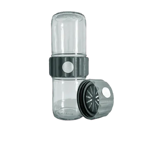
BioCoupler® Glass Set (16 oz) x12
BCS1612
12 Each
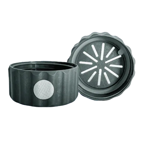
BioCoupler® x 48
BC48
48 Each
BioTilt™ 25 (BT25)
BT25
25 Bundles
Purified Diphtheria Toxoid protein (antigen grade)
DTOX15-N-500
100 µg
Recent gezochte producten
SMRCD Polyclonal Antibody
JOT-AP14328-01
20 µL
MYCD Polyclonal Antibody
JOT-AP11603-01
20 µL
Anti-DCD Polyclonal Antibody
HX130014-01
50 µg
Anti-MYOCD Polyclonal Antibody
HV334024-01
50 µg
Anti-Human FTCD Polyclonal Antibody
HF652014-01
50 µg
Anti-Human PIK3CD Polyclonal Antibody
HT068014-01
50 µg
Anti-Human PRCD Polyclonal Antibody
HD164014-01
50 µg
SCD polyclonal antibody
GTR18043932-01
50 μL