

Humaan Arginase 2/ARG2-antilichaam
Kwaliteit
ISO Gecertificeerd
Levering
24-48 uur
Technische Specificaties
Arginase 2 catalyzes the same reaction as arginase 1, but differs in its tissue specificity and subcellular location. Specifically, arginase 2 localizes to the mitochondria. Arginase 2 is expressed in non-hepatic tissues, with the highest levels of expression in the kidneys, but, unlike arginase 1, is not expressed in liver.
Arginase-2 type II, Kidney arginase, Non-hepatic arginase, Arginase-2, type II Arginase II mitochondrial, Kidney type arginase, Non hepatic arginase.
• Mouse
Human
Human
Recombinant human ARG2 (23-354aa) purified from E. coli
• Monoclonal
• IgG1 κ
AT7F8
Unconjugated
ELISA, WB, ICC/IF, FACS
By protein-A affinity chromatography
1 mg/mL (determined by BCA assay)
Arginase2, ARG2, Arginase-2 type II, Kidney arginase, Non-hepatic arginase, Arginase-2, type II Arginase II mitochondrial, Kidney type arginase, Non hepatic arginase., ATGA0389-10 µg, ATGA0389-20 µg, ATGA0389-50 µg, ATGA0389-100 µg, ATGA0389-250 µg, ATGA0389-500 µg, ATGA0389-1 mg, ATGA0389-10, ATGA0389-20, ATGA0389-50, ATGA0389-100, ATGA0389-250, ATGA0389-500, ATGA0389-1
Watts JA., et al. (2011) Pulm Pharmacol Ther. 24 (4) : 407-413.; ; Tadie JM., et al. (2008) Am J Physiol Lung Cell Mol Physiol. 294 (3) : 489-497.
Can be stored at 2°C to 8°C for 1 week. For long term storage, aliquot and store at -20C to -80C. Avoid repeated freezing and thawing cycles.
Liquid in. Phosphate-Buffered Saline (pH 7.4) with 0.02% Sodium Azide, 10% glycerol
The antibody has been tested by ELISA, Western blot analysis, Flow cytometry and ICC/IF to assure specificity and reactivity. Since application varies, however, each investigation should be titrated by the reagent to obtain optimal results.
Metabolism
NP_001163
P78540
The SW480 Cell lysate (40ug) was resolved by SDS-PAGE, transferred to PVDF membrane and probed with anti-human ARG2 antibody (1:1000) . Proteins were visualized using a goat anti-mouse secondary antibody conjugated to HRP and an ECL detection system. ; ; Lane 1. : SW480 cell lysate The recombinant proteins (50ng) were resolved by SDS-PAGE, transferred to PVDF membrane and probed with anti-human ARG2 antibody (1:1000) . Proteins were visualized using a goat anti-mouse secondary antibody conjugated to HRP and an ECL detection system.; ; Lane 1.: ARG2 recombinant protein (ATGP0591) ; ; Lane 2.: ARG1 recombinant protein (ATGP0564) {2004010328382040.jpg}
Flow cytometry analysis of ARG2 in SW480 cell line, staining at 2-5ug for 1x10⁶cells (red line) . The secondary antibody used goat anti-mouse IgG Alexa fluor 488 conjugate. Isotype control antibody was mouse IgG (black line) .
ICC/IF analysis of ARG2 in SW480 cells line, stained with DAPI (Blue) for nucleus staining and monoclonal anti-human ARG2 antibody (1:100) with goat anti-mouse IgG-Alexa fluor 488 conjugate (Green) .
Beschrijving
Arginase 2 catalyzes the same reaction as arginase 1, but differs in its tissue specificity and subcellular location. Specifically, arginase 2 localizes to the
Specificaties
Recent bekeken producten
SCD polyclonal antibody
GTR18043932-01
50 μL
(KO Validated) SUMO1 Polyclonal Antibody
E-AB-92410-01
60 µL
Salicylate hydroxylase, Microorganism
HY-E70520-01
1 KU
Purified Diphtheria Toxoid protein (cGMP/USP, vaccine grade, ~1500-2500 Lf/ml; Low endotoxin)
DTOX16-N-500
500 µg
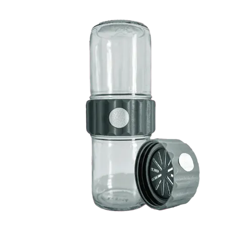
BioCoupler® Glass Set (16 oz) x12
BCS1612
12 Each
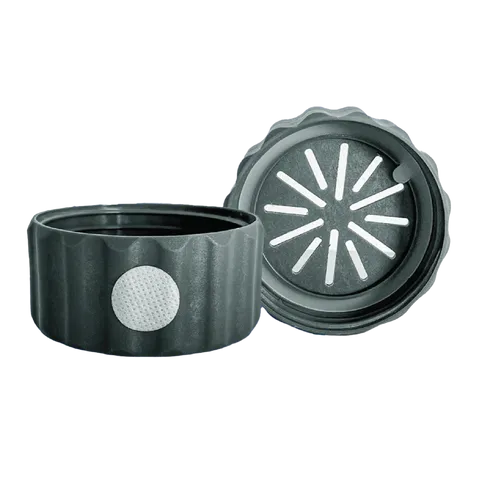
BioCoupler® x 48
BC48
48 Each
BioTilt™ 25 (BT25)
BT25
25 Bundles
Purified Diphtheria Toxoid protein (antigen grade)
DTOX15-N-500
100 µg
Recent gezochte producten
SMRCD Polyclonal Antibody
JOT-AP14328-01
20 µL
MYCD Polyclonal Antibody
JOT-AP11603-01
20 µL
Anti-DCD Polyclonal Antibody
HX130014-01
50 µg
Anti-MYOCD Polyclonal Antibody
HV334024-01
50 µg
Anti-Human FTCD Polyclonal Antibody
HF652014-01
50 µg
Anti-Human PIK3CD Polyclonal Antibody
HT068014-01
50 µg
Anti-Human PRCD Polyclonal Antibody
HD164014-01
50 µg
SCD polyclonal antibody
GTR18043932-01
50 μL