

Humaan IRF3-antilichaam
Kwaliteit
ISO Gecertificeerd
Levering
24-48 uur
Technische Specificaties
Members of the Interferon regulatory factor (IRF) family regulate gene expression critical to immune response, hemopoiesis, and proliferation. IRF-3 is a member of the IRF family, and is distinct from other family members. Its transcriptional activity is regulated solely by posttranslational modifications. It plays a crucial role in activation of innate immunity and inflammation in response to viral infection.
IRF3, Interferon regulatory factor-3, IRF-3, Interferon regulatory factor 3, IRF 3
• Mouse
Human
Human
Recombinant human IRF3 (108-166aa) purified from E. coli
• Monoclonal
• IgG1 κ
3F10
Unconjugated
ELISA, WB, ICC/IF, FACS
By protein-G affinity chromatography
1 mg/mL (determined by BCA assay)
IRF3, AIR0718-10 µg, AIR0718-20 µg, AIR0718-50 µg, AIR0718-100 µg, AIR0718-250 µg, AIR0718-500 µg, AIR0718-1 mg, AIR0718-10, AIR0718-20, AIR0718-50, AIR0718-100, AIR0718-250, AIR0718-500, AIR0718-1
DeFilippis VR et al., (2006) J Virol 80 (2) :1032-1037.; ; Pietila TE et al., (2007) J Immunol 178 (1) :253-261.
Can be stored at 2°C to 8°C for 1 week. For long term storage, aliquot and store at -20C to -80C. Avoid repeated freezing and thawing cycles.
Liquid in. Phosphate-Buffered Saline (pH 7.4) with 0.02% Sodium Azide, 10% glycerol
The antibody has been tested by ELISA, Western blot, ICC/IF and FACS analysis to assure specificity and reactivity. Since application varies, however, each investigation should be titrated by the reagent to obtain optimal results.
Immunology, Hormones & Cytokines
NP_001562
Q14653
The cell lysates (40ug) were resolved by SDS-PAGE, transferred to PVDF membrane and probed with anti-human IRF3 antibody (1:1000) . Proteins were visualized using a goat anti-mouse secondary antibody conjugated to HRP and an ECL detection system.; ; Lane 1.: MCF7 cell lysate; ; Lane 2.: K562 cell lysate; ; Lane 3.: Jurkat cell lysate
Flow cytometry analysis of IRF3 in MCF7 cells. The cell was stained with AIR0718 at 2-5ug for 1x10^6cells (red) . A Goat anti mouse IgG (Alexa fluor 488) was used as the secondary antibody. Mouse monoclonal IgG was used as the isotype control (dark gray), cells without incubation with primary and secondary antibody was used as the negative control (light gray) . Flow cytometry analysis of IRF3 in Jurkat cells. The cell was stained with AIR0718 at 2-5ug for 1x10^6cells (red) . A Goat anti mouse IgG (Alexa fluor 488) was used as the secondary antibody. Mouse monoclonal IgG was used as the isotype control (dark gray), cells without incubation with primary and secondary antibody was used as the negative control (light gray) . {2004010048549707.jpg}
ICC/IF analysis of human HeLa cells stained with IRF3 (1:500) with Texas-Red and Hoechst 33342 (Blue) for nucleus staining.
Beschrijving
Members of the Interferon regulatory factor (IRF) family regulate gene expression critical to immune response, hemopoiesis, and proliferation. IRF-3 is a member
Specificaties
Recent bekeken producten
AGPAT4 Antibody
MBS7124622-01
0.05 mL
H&E Mount, Permanent Mountindg media for coverslipping H&E stains from water,
NB315
125 mL
SCD polyclonal antibody
GTR18043932-01
50 μL
(KO Validated) SUMO1 Polyclonal Antibody
E-AB-92410-01
60 µL
Salicylate hydroxylase, Microorganism
HY-E70520-01
1 KU
Purified Diphtheria Toxoid protein (cGMP/USP, vaccine grade, ~1500-2500 Lf/ml; Low endotoxin)
DTOX16-N-500
500 µg
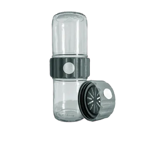
BioCoupler® Glass Set (16 oz) x12
BCS1612
12 Each
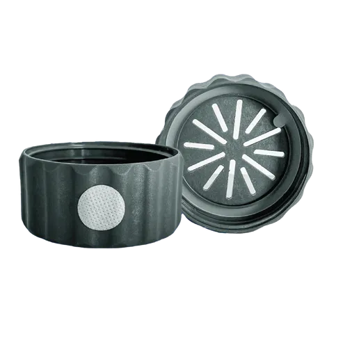
BioCoupler® x 48
BC48
48 Each