

Menselijk EDAR antilichaam
Kwaliteit
ISO Gecertificeerd
Levering
24-48 uur
Technische Specificaties
Tumor necrosis factor receptor superfamily member EDAR is a protein that in humans is encoded by the EDAR gene. EDAR and other genes provide instructions for making proteins that work together during embryonic development. These proteins form part of a signaling pathway that is critical for the interaction between two cell layers, the ectoderm and the mesoderm. In the early embryo, these cell layers form the basis for many of the body's organs and tissues. Ectoderm-mesoderm interactions are essential for the proper formation of several structures that arise from the ectoderm, including the skin, hair, nails, teeth, and sweat glands.
Tumor necrosis factor receptor superfamily member EDAR, DL, ECTD10A, ECTD10B, ED1R, ED3, ED5, EDA-A1R, EDA1R, EDA3, HRM1
• Mouse
Human
Human
Recombinant human EDAR (27-448aa) purified from E.coli
• Monoclonal
• IgG2a κ
AT19E8
Unconjugated
ELISA, WB, FACS
By protein-A affinity chromatography
1 mg/mL (determined by BCA assay)
EDAR, ATGA0334-10 µg, ATGA0334-20 µg, ATGA0334-50 µg, ATGA0334-100 µg, ATGA0334-250 µg, ATGA0334-500 µg, ATGA0334-1 mg, ATGA0334-10, ATGA0334-20, ATGA0334-50, ATGA0334-100, ATGA0334-250, ATGA0334-500, ATGA0334-1
Monreal AW, et al. (1999) Nat Genet 22 (4) :366-9.; ; Aswegan AL, et al. (1997) Am J Med Genet 72 (4) :462-7.; ; Entrez Gene: EDAR ectodysplasin A receptor.
Can be stored at 2°C to 8°C for 1 week. For long term storage, aliquot and store at -20C to -80C. Avoid repeated freezing and thawing cycles.
Liquid in. Phosphate-Buffered Saline (pH 7.4) with 0.02% Sodium Azide, 10% glycerol
The antibody has been tested by ELISA, Western blot and FACS analysis to assure specificity and reactivity. Since application varies, however, each investigation should be titrated by the reagent to obtain optimal results.
Signal Transduction
NP_071731
Q9UNE0
The recombinant protein (50ng) and cell lysates (40ug) were resolved by SDS-PAGE, transferred to PVDF membrane and probed with anti-human EDAR antibody (1:1000) . Proteins were visualized using a goat anti-mouse secondary antibody conjugated to HRP and an ECL detection system. ; ; Lane 1 : Recombinant protein EDAR ; ; Lane 2 : A549 cell lysate ; ; Lane 3 : Ramos cell lysate
Flow cytometry analysis of EDAR in HeLa cells. The cell was stained with ATGA0334 at 2-5ug for 1x10^6cells (red) . A Goat anti mouse IgG (Alexa fluor 488) was used as the secondary antibody. Mouse monoclonal IgG was used as the isotype control (dark gray), cells without incubation with primary and secondary antibody was used as the negative control (light gray)
Beschrijving
Tumor necrosis factor receptor superfamily member EDAR is a protein that in humans is encoded by the EDAR gene. EDAR and other genes provide instructions for ma
Specificaties
Recent bekeken producten
AGPAT4 Antibody
MBS7124622-01
0.05 mL
H&E Mount, Permanent Mountindg media for coverslipping H&E stains from water,
NB315
125 mL
SCD polyclonal antibody
GTR18043932-01
50 μL
(KO Validated) SUMO1 Polyclonal Antibody
E-AB-92410-01
60 µL
Salicylate hydroxylase, Microorganism
HY-E70520-01
1 KU
Purified Diphtheria Toxoid protein (cGMP/USP, vaccine grade, ~1500-2500 Lf/ml; Low endotoxin)
DTOX16-N-500
500 µg
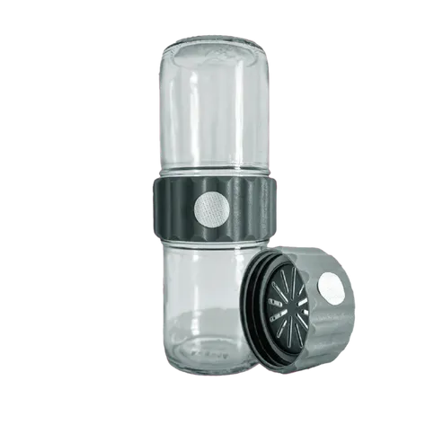
BioCoupler® Glass Set (16 oz) x12
BCS1612
12 Each
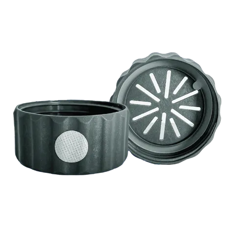
BioCoupler® x 48
BC48
48 Each