

Menselijk OSTF1-antilichaam
Kwaliteit
ISO Gecertificeerd
Levering
24-48 uur
Technische Specificaties
Osteoclast-stimulating factor (OSTF1) is an intracellular signaling protein, highly expressed by osteoclasts (OCL) . Indirectly, it enhances osteoclast (OCL) formation and bone resorption through a cellular signal transduction cascade by possibly interacting with c-Src or other c-Src-related proteins. OSTF1 contains a short proline-rich N-terminal region, an SH3 domain, three ankyrin repeats and an aspartate-rich C-terminal region, suggesting that it is potentially involved in protein-protein interactions.
Osteoclast stimulating factor 1, SH3P2, OSF, Osteoclast stimulating factor 1
• Mouse
Human
Human
Recombinant human OSTF1 (1-217aa) purified from E. coli
• Monoclonal
• IgG2a κ
AT9G4
Unconjugated
ELISA, WB, ICC/IF, FACS
By protein-G affinity chromatography
1 mg/mL (determined by BCA assay)
OSTF1, ATGA0125-10 µg, ATGA0125-20 µg, ATGA0125-50 µg, ATGA0125-100 µg, ATGA0125-250 µg, ATGA0125-500 µg, ATGA0125-1 mg, ATGA0125-10, ATGA0125-20, ATGA0125-50, ATGA0125-100, ATGA0125-250, ATGA0125-500, ATGA0125-1
Kurihara N., et al. (2001) J. Biol. Chem. 276 (44) : 41035-9. ; ; Reddy S., et al. (1999) J Cell Physiol. 177 (4) : 636-45.
Can be stored at 2°C to 8°C for 1 week. For long term storage, aliquot and store at -20C to -80C. Avoid repeated freezing and thawing cycles.
Liquid in. Phosphate-Buffered Saline (pH 7.4) with 0.02% Sodium Azide, 10% glycerol
The antibody has been tested by ELISA, Western blot, ICC/IF and FACS analysis to assure specificity and reactivity. Since application varies, however, each investigation should be titrated by the reagent to obtain optimal results.
Metabolism
AAH07459
Q92882
The cell lysates (40ug) were resolved by SDS-PAGE, transferred to PVDF membrane and probed with anti-human OSTF1 antibody (1:1000) . Proteins were visualized using a goat anti-mouse secondary antibody conjugated to HRP and an ECL detection system.; ; Lane 1.: HepG2 cell lysate; ; Lane 2.: MCF7 cell lysate; ; Lane 3.: Jurkat cell lysate; ; Lane 4.: LnCap cell lysate The cell lysates (10 µg) were resolved by SDS-PAGE, transferred to PVDF membrane and probed with anti-human OSTF1 antibody (1:1000) . Proteins were visualized using a goat anti-mouse secondary antibody conjugated to HRP and an ECL detection system.; ; Lane 1.: 293T cell lysate; ; Lane 2.: OSTF1 Transfected 293T cell lysate {ATGA0125-WB2.jpg}
Flow cytometry analysis of OSTF1 in HeLa cell line, staining at 2-5ug for 1x10^6cells (red line) . The secondary antibody used goat anti-mouse IgG Alexa fluor 488 conjugate. Isotype control antibody was mouse IgG (black line) .
ICC/IF analysis of OSTF1 in MCF7 cells. The cell was stained with ATGA0125 (1:100) . The secondary antibody (green) was used Alexa Fluor 488. DAPI was stained the cell nucleus (blue) .ICC/IF analysis of OSTF1 in A431 cells. The cell was stained with ATGA0125 (1:100) . The secondary antibody (green) was used Alexa Fluor 488. DAPI was stained the cell nucleus (blue) .{ATGA0125-IF2.jpg}
Beschrijving
Osteoclast-stimulating factor (OSTF1) is an intracellular signaling protein, highly expressed by osteoclasts (OCL) . Indirectly, it enhances osteoclast (OCL) fo
Specificaties
Recent bekeken producten
Arachis hypogaea Lectin (PNA) - Biotinylated
21510020-1
5mg
1,4- Dioxane Pure
1041330 500
500 mL
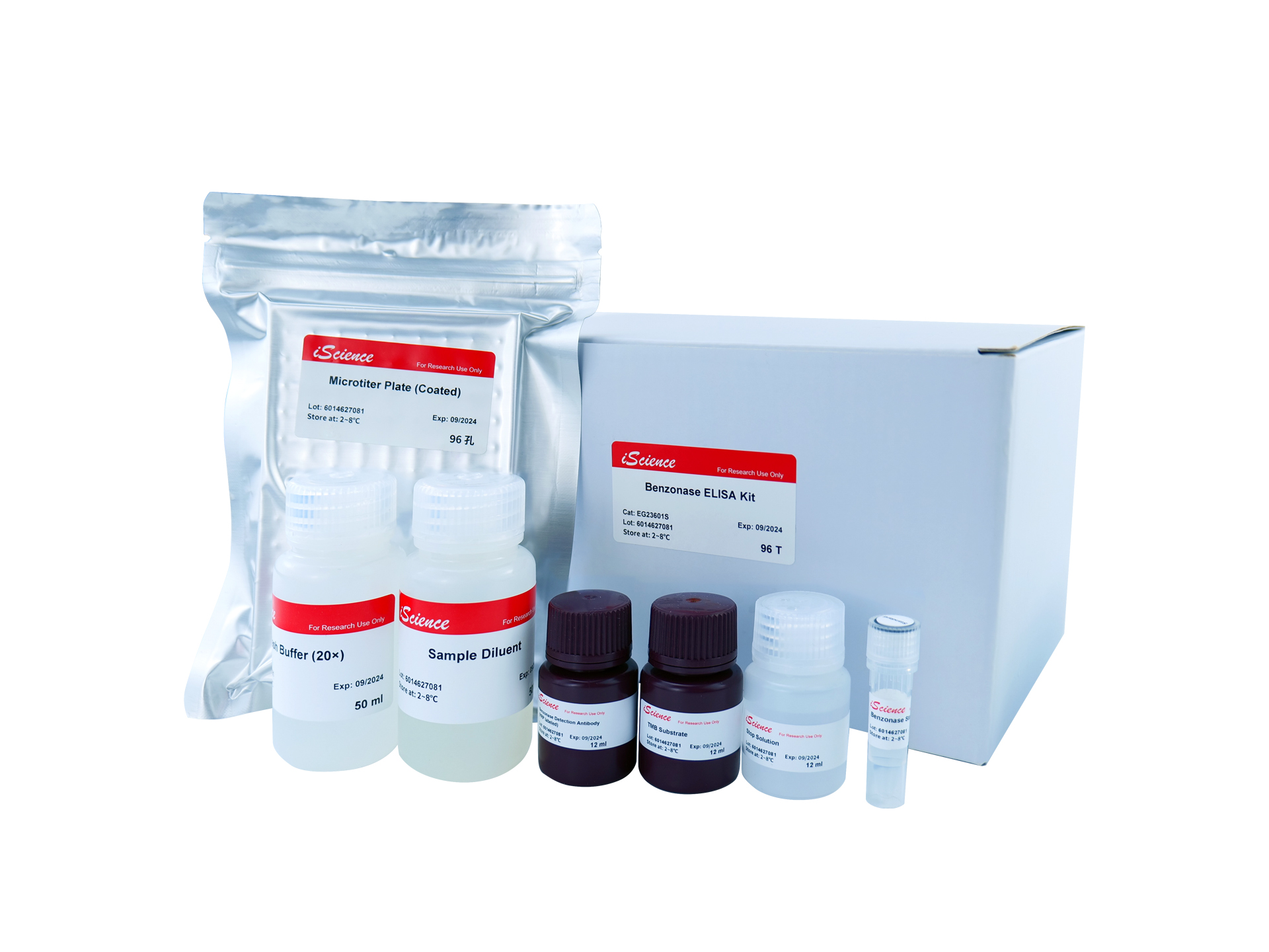
Benzonase ELISA Kit
EG23601S
96 Tests

Not I Restriction Enzyme
B2016623
500 Units
K562-HLA-G0101 Overexpressing Cell Line
RG-1214
5x 10^6
Yeast Poly (A) Polymerase
RNT-006-S
30000 Units
Arachis hypogaea lectin (PNA)
05-0116-10mg
10 mg
Anti-APOE2 mouse monoclonal antibody
TD-AB0132
100 µg