

PHAP I Antilichaam
Kwaliteit
ISO Gecertificeerd
Levering
24-48 uur
Technische Specificaties
PHAP I Antibody: Apoptosis is related to many diseases and development. Caspase-9 plays a central role in cell death induced by a variety of apoptosis activators. Cytochrome c, after released from mitochondria, binds to Apaf-1, which forms an apoptosome that in turn binds to and activate procaspase-9. Activated caspase-9 cleaves and activates the effector caspases (caspase-3, -6 and -7), which are responsible for the proteolytic cleavage of many key proteins in apoptosis. The tumor suppressor putative HLA-DR-associated proteins (PHAPs) were recently identified as important regulators of mitochondrion apoptosis. PHAP appears to facilitate apoptosome-medicated caspase-9 activation and to stimulate the mitochondrial apoptotic pathway. PHAP was also shown to oppose both Ras- and Myc-medicated cell transformation.
8125
P39687
P39687
• Rabbit
Human, Mouse, Rat
3145
• Polyclonal
Unconjugated
Primary Antibodies
Independent Antibody Validation in Cell lines (Figure 2) shows similar PHAP I expression profile in human, mouse and rat cell lines detected by two independent anti-PHAP I antibodies that recognize different epitopes, 3145 against C-terminus domain and 3151 against C-terminus different domain. PHAP I proteins are detected in the most tested cell lines at different expression levels by the two independent antibodies. Independent Antibody Validation in Cell lines3145 3151 KD validation (Figure 6) : Anti-PHAP I antibody (3151) specificity was further verified by PHAP I specific knockdown. PHAP I signal in T47D cells transfected with PHAP I siRNAs was disrupted in comparison with that in cells transfected with control siRNAs. KD validation3151 Regulated expression validation (Figure 7, 8, 9) : PHAP I expression detected by anit-PHAP I antibodies (3151) was up-regulated in breast cancer tissues of patients (Figure 7) and the breast cancer cell line (Figure 8) as well as in the Atxn1 KO mice (Figure 9) . Regulated expression validation3151
Apoptosis
PHAP I Antibody is DEAE purified.
Cat. No. 1207 - Raji Cell Lysate
1 mg/mL
PHAP I Antibody is supplied in PBS containing 0.02% sodium azide.
None
Blue Ice
PHAP I antibody can be stored at 4˚ C for three months and -20˚ C, stable for up to one year. As with all antibodies care should be taken to avoid repeated freeze thaw cycles. Antibodies should not be exposed to prolonged high temperatures.
IgG
This polyclonal antibody has no cross-reaction to PHAP I2a and PHAP III.
Human PHAP I has one isoform (249aa, 28.6kD) . Mouse PHAP I has one isoform (247aa, 28.5kD) and rat PHAP I also has one isoform (247aa, 28.6kD) . 3151 can detect all isoforms of human, mouse and rat.
ANP32A
Cat. No. 17-007 - Raji Cell Slide
Acidic (leucine-rich) nuclear phosphoprotein 32 family, member A
Homo sapiens
Jiang et al. Science. 2003;299 (5604) :223-6.
Nicholson and Thornberry. Science. 2003 10;299 (5604) :214-5.
16489023
23630944
23652204
Parrish. Regulation of Apoptosis Following Mitochondrial Cytochrome c Release. Department of Pharmacology and Molecular Cancer Biology, Duke University. PhD Thesis, 2010.
PHAP I Antibody: LANP, MAPM, PP32, HPPCn, PHAP1, PHAPI, I1PP2A, C15orf1, LANP, Acidic leucine-rich nuclear phosphoprotein 32 family member A, Acidic nuclear phosphoprotein pp32
ELISA, WB, ICC, IF
730318
Liquid
Beschrijving
PHAP I Antilichaam Beschikbaar in 0.02 mg. Bestel eenvoudig online met snelle levering.
Specificaties
Recent bekeken producten
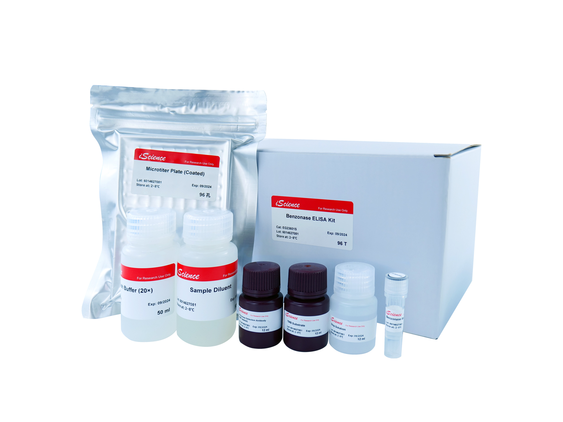
Benzonase ELISA Kit
EG23601S
96 Tests

Not I Restriction Enzyme
B2016623
500 Units
K562-HLA-G0101 Overexpressing Cell Line
RG-1214
5x 10^6
Yeast Poly (A) Polymerase
RNT-006-S
30000 Units
Arachis hypogaea Lectin (PNA) - Biotinylated
21510020-1
5mg
Arachis hypogaea lectin (PNA)
05-0116-10mg
10 mg
Anti-APOE2 mouse monoclonal antibody
TD-AB0132
100 µg
Human Apolipoprotein E2 (Apo E2) Antibody
22210-05011
150 µg