

Bijlagein V-FITC Apoptosis Detection Kit
Kwaliteit
ISO Gecertificeerd
Levering
24-48 uur
Technische Specificaties
Apoptosis or programmed cell death is an evolutionary conserved, intrinsic program of cell death that occurs in various physiological and pathological situations. It is a key regulator of physiological growth control and regulation of tissue homeostasis. Apoptosis is considered a vital component of various processes including normal cell turnover, proper development and functioning of the immune system, hormone-dependent atrophy, embryonic development and chemical-induced cell death. The term apoptosis (a-po-toe-sis) was first used in a now-classic paper by Kerr, Wyllie, and Currie in 1972 to describe a morphologically distinct form of cell death, although certain components of the apoptosis concept had been explicitly described many years previously. Apoptosis is characterized by specific morphological changes of the dying cells; namely, loss of membrane asymmetry, cytoskeleton remodeling, plasma membrane blebbing, loss of the mitochondrial membrane potential, caspase activation, chromatin condensation, and DNA fragmentation. During apoptosis, the asymmetric distribution of phospholipids of the plasma membrane gets lost and phosphatidylserine (PS) is translocated to the outer leaflet of the plasma membrane. There, PS acts as one major “eat me†signal that ensures efficient recognition and uptake of apoptotic cells by phagocytes. Once on the cell surface, PS can be easily detected by staining with a fluorescent conjugate of Annexin V, a 35-36 kDa Ca2+ dependent phospholipid-binding protein that has a high affinity for PS, and binds to cells with exposed PS. The one-step staining procedure takes only 10 minutes. Detection can be analyzed by flow cytometry or by fluorescence microscopy. Annexin V may be conjugated to fluorochromes including FITC. This format retains its high affinity for PS and thus serves as a sensitive probe for flow cytometric analysis of cells that are undergoing apoptosis.
FACS
Store all kit components at 4°C.
STAINING PROTOCOL 1. Before beginning of the experiment, please dilute 10X PBS and 10X Binding Buffer to respective 1X by using distilled water.Please label the vials as follows (Table 1) . 2. Control cells should be re-suspended properly by gently tapping the tube before initiation of staining. Aliquot 100μl of control cells to 3 individual vials as indicated in the Table 1 (Tube no 1, 2 and 3) . 3. Resuspend the cells with 1 ml of pre-cooled PBS. Centrifuge the cells at 300 x g for 5 min. 4. Decant the supernatant carefully and repeat the washing step 6. After decanting the supernatant, resuspend the pellet with 100 μl 1X Binding Buffer by gently tapping the tube. DO NOT VORTEX. 7. Wash the experimental cells twice with 1 ml of pre-cooled 1X PBS by centrifuge the cells at 300 x g for 5 min. Resuspend the pellet in (~1 x 10⁶ cells/ml) 1X Binding Buffer. 8. Aliquot 100 μl of Experimental Cells to tubes no 4 and 5 as indicated in Table 1. 9. Add 5 μl of the Staining Reagents to each tube as indicated in Table 1 and mix the cells suspension thoroughly. 10. Incubate for 20 min at RT (20-25°C) in the dark. After incubation add 400 μl of 1X Binding Buffer to each tube. 11. Samples should be analyzed within 1 hr of the staining. Data acquisition should be done in FL1 channel for detecting Annexin V- FITC staining and the FL2 channel for detecting PI staining.
Beschrijving
Apoptosis or programmed cell death is an evolutionary conserved, intrinsic program of cell death that occurs in various physiological and pathological situation
Specificaties
Recent bekeken producten
Anti-TAT (Thrombin-antithrombin) Antibody
ABS 014-22-1
1000 µL
Anti-APOE2 mouse monoclonal antibody
TD-AB0132
100 µg
CD35 (CR1) mouse monoclonal antibody, clone J3D3, Azide Free
BM2364
0.2 mg
Anti-KIR2DL1 (Rabbit, Monoclonal, Rabbit mAb)
A115392
100 µL
Stains-all
03943-5
5 g
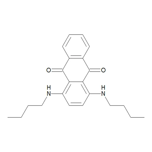
Sudan Blue II;Oil Blue 35
TRC-S688913-500MG
500 mg
Nile Blue A
N8430-01
1 g
Human Apolipoprotein E2 (Apo E2) Antibody
22210-05011
150 µg
Recent gezochte producten
QuickAntibody Mouse2W (2-week standard mouse polyclonal antibody preparation adjuvant)
E28X0210043
1 mL
Mouse Polyclonal Nanos2 Antibody
H00339345-B01P
50 µg
CBS polyclonal antibody
BS6545-01
50 µL
CBS polyclonal antibody
BS90205
50ul
EPO polyclonal antibody
GTR18043715-01
50 μL
SHB polyclonal antibody
GTR18213369-01
50 μL
SHB polyclonal antibody
GTR18291465-01
50 µL
CSTB polyclonal antibody
GTR18295782-01
50 µL