

BLOW IntraCellular Staining Kit (IC FLOW)
Kwaliteit
ISO Gecertificeerd
Levering
24-48 uur
Technische Specificaties
The Intracellular FLOW Staining Kit allows detection antibodies to access the intracellular protein in Flowcytometry analysis. This kit is specially formulated to reduce non-specific staining of fluorochrome-labelled antibodies and to increase fluorescence signaling. First, live cells can be fixed with the Fixation buffer, which cross-links the protein. Second, the 1X Permeabilization Buffer creates holes in the membrane, allowing the antibodies enter into the cells effectively. Washing steps and antibody dilutions are done using 1X Permeabilization Buffer. The final cell resuspension prior to analysis is done using The Staining Buffer. Permeabilization Buffer is supplied as a 10X solution. Prior to use, this should be diluted 10 fold in distilled water. (1ml of Permeabilization Buffer with 9 ml of distilled water) . 10X Permeabilization buffer has natural tendency to precipitate, however, its function is not affected. 1X solution can be filtered.
FACS
Kit Components: Fixation Buffer (1X) - 60 ml, Permeabilization Buffer (10X) - 2 X 60 ml, Staining Buffer - (1X) - 60 ml
Store at 2-8°C
Protocol: 1. Determine number of cells required for staining. Each sample contains 0.5 -1 x 10^6 cells in 50μl of media or staining buffer. The following controls are needed for the experiment. Unstained cells (no antibodies were added), cells with isotype control and cells with secondary antibody (if secondary antibody was used) . 2. Cetrifuge cells at 1000 RPM for 10 minutes and decant supernatant. 3. Resuspend cells with appropriate volume of Fixation buffer. Incubate in ice for for 30 minutes. 4. Centrifuge cells 1000 RPM for 10 minutes and decant supernatant. 5. Resuspend pellet with appropriate volume of 1X permeabilization buffer. 6. Aliquote 1x10^6 cells in 50 μl to the desired number of flow tubes. Dilute primary antibodies in 50 μl of 1X permeabilization buffer. Add diluted antibodies into 50 μl of cells. Mix antibodies in cells suspension thoroughly. 7. Incubate in ice for 30 minutes. Wash cells in 2-3 ml of 1X permeabilization buffer. Centrifuge 1000 RPM for 10 minutes. Decant supernatant carefully. If primary antibody is fluorescent-labeled, resuspent pellet in 300-400 μl of staining buffer, analyze samples. Protected from light. If primary antibody is not labeled, proceed with step 8. 8. Dilute secondary antibody (PE, FITC, APC or biotin labeled) in 50 μl of permeabilization buffer per sample. 9. Resuspend cells in diluted secondary antibody. Incubate in ice for 30 minutes. (protected from light) . 10. Wash cells in 3-4 ml of permeabilization buffer. Cetrifuge at 1000 RPM for 10 minutes. Decant supernatant. 11. After decanting, add 300-400 μl of Staining Buffer to each tube. If not analyzing same day, samples can be stored over night in dark at 4 degrees C. Samples can be analyzed in Flow Cytometer according to the manufacturer protocol.
Beschrijving
The Intracellular FLOW Staining Kit allows detection antibodies to access the intracellular protein in Flowcytometry analysis. This kit is specially formulated
Specificaties
Recent bekeken producten
Anti-TAT (Thrombin-antithrombin) Antibody
ABS 014-22-1
1000 µL
Anti-APOE2 mouse monoclonal antibody
TD-AB0132
100 µg
CD35 (CR1) mouse monoclonal antibody, clone J3D3, Azide Free
BM2364
0.2 mg
Anti-KIR2DL1 (Rabbit, Monoclonal, Rabbit mAb)
A115392
100 µL
Stains-all
03943-5
5 g
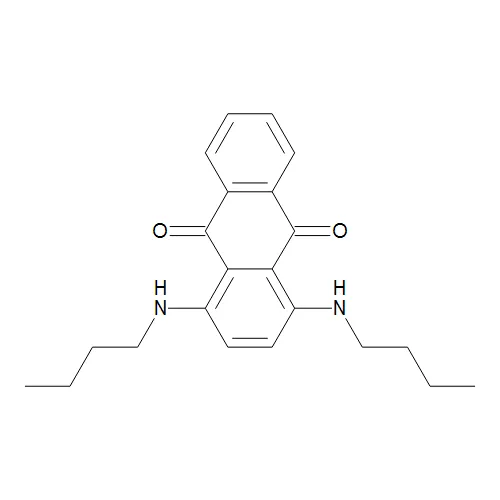
Sudan Blue II;Oil Blue 35
TRC-S688913-500MG
500 mg
Nile Blue A
N8430-01
1 g
Human Apolipoprotein E2 (Apo E2) Antibody
22210-05011
150 µg
Recent gezochte producten
QuickAntibody Mouse2W (2-week standard mouse polyclonal antibody preparation adjuvant)
E28X0210043
1 mL
Mouse Polyclonal Nanos2 Antibody
H00339345-B01P
50 µg
CBS polyclonal antibody
BS6545-01
50 µL
CBS polyclonal antibody
BS90205
50ul
EPO polyclonal antibody
GTR18043715-01
50 μL
SHB polyclonal antibody
GTR18213369-01
50 μL
SHB polyclonal antibody
GTR18291465-01
50 µL
CSTB polyclonal antibody
GTR18295782-01
50 µL